
Draw a neat labelled diagram of monocot stem.
Anatomy of Monocot Root Cross Section The anatomical features of a monocot root can be studied through a cross section (CS) through the root. The present post discesses the anatomy of monocot root with Monocot Root Diagram. Ø Anatomically, the monocot root has been differentiated into the following parts: (1). Epidermis (2). Cortex (3). Endodermis

Monocot Stem Cross Section Monocot Root Cross Section Structure (with PPT The main
Organ-specific vascular tissue organisation in the monocot rice and the dicot Arabidopsis. (a) Transverse section through a mature rice root. Vascular tissues are organised in a central vascular cylinder surrounded by the endodermis and embedded in the cortex. (b) Transverse section through a mature rice stem internode.
Chapter 9 Transport in Plants Lesson 1 Monocotyledons and dicot
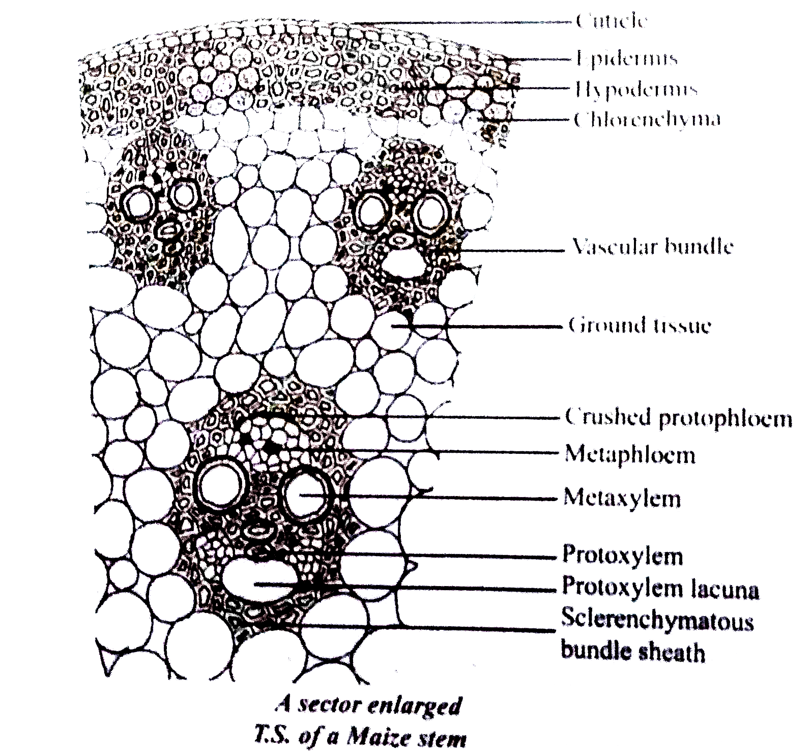
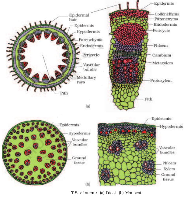
Preparation and Study of the Transverse Section of Dicot and Monocot Roots and Stems Dicot stem Aim The aim of this study is to closely observe the sentient transverse dicot stem. Requirements Some essential materials are required like root as well as stem of a sunflower, fresh root as well as stem of maize, a microscope as well as the blade.
Monocot Stem Cross Section
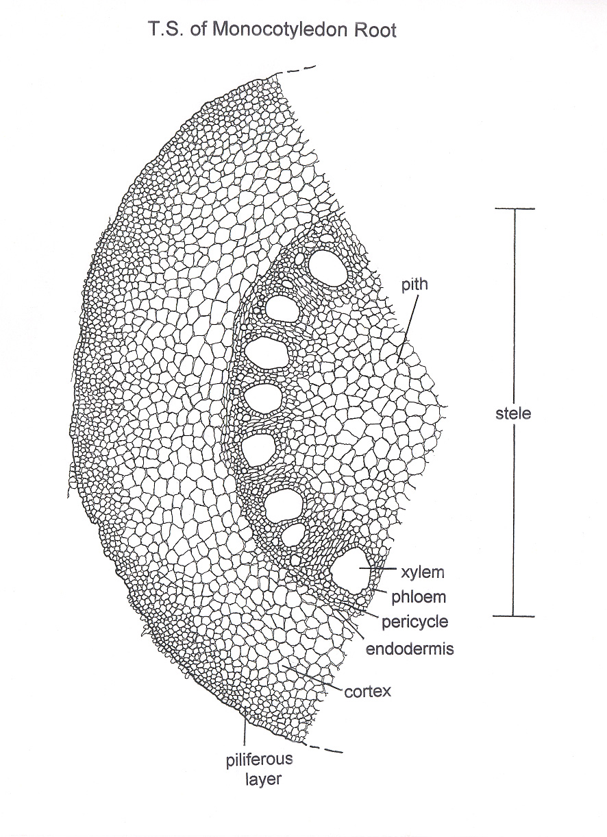
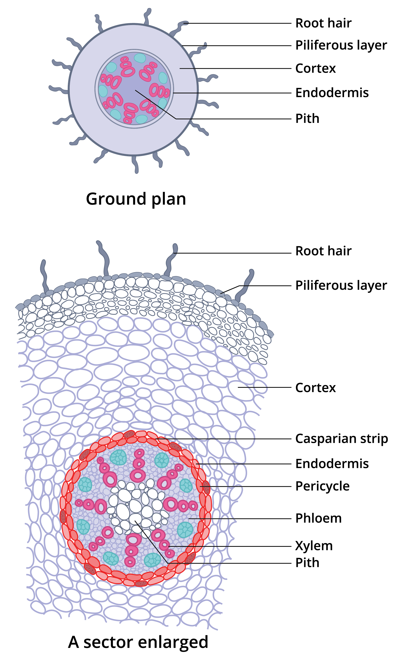
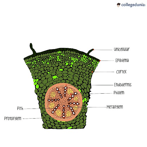
The transverse section of the monocot root (maize) shows the following plan of arrangement of tissues from the periphery to the centre. Piliferous Layer or Epiblema. The outermost layer of the root is known as piliferous layer. It consists of a single row of thin-walled parenchymatous cells without any intercellular space. Epidermal pores and.

Draw and label the parts of transverse section of monocot root. Brainly.in
The dicot roots are covered by exodermis which is a modified epidermis. Cortex. The cortex in monocot roots is wide. The cortex in dicot roots is narrow. The cortex in monocot roots is composed of only parenchymatous cells. The cortex of dicot roots is composed of both parenchymatous and sclerenchymatous cells.
Micscape Image Gallery Botanical Drawing
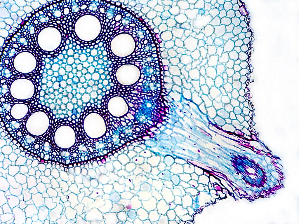
Monocot Root Anatomy PPT: Anatomy of Monocot Root, Monocot Root Transverse Section Anatomical Details, TS Monocot Root under the Microscope, Record Diagram for Monocot Root, Epidermis, Cortex, Endodermis and Stele of Monocot Roots, Stelar Features of Monocot Roots, Learn more: Notes on Anatomy of Monocot Roots
class on structure of monocot stem YouTube
Anatomy of monocot root: In many aspects, it is similar to the dicot root. On comparing the two roots, we find the monocot root also consists of epidermis, cortex, endodermis, pericycle, vascular bundle and pith.. Going back to the transverse section of the dicot root, after the endodermis, a few layers of thick-walled parenchymatous cells.
Internal structure of monocot root (Maize) — lesson. Science State Board, Class 10.
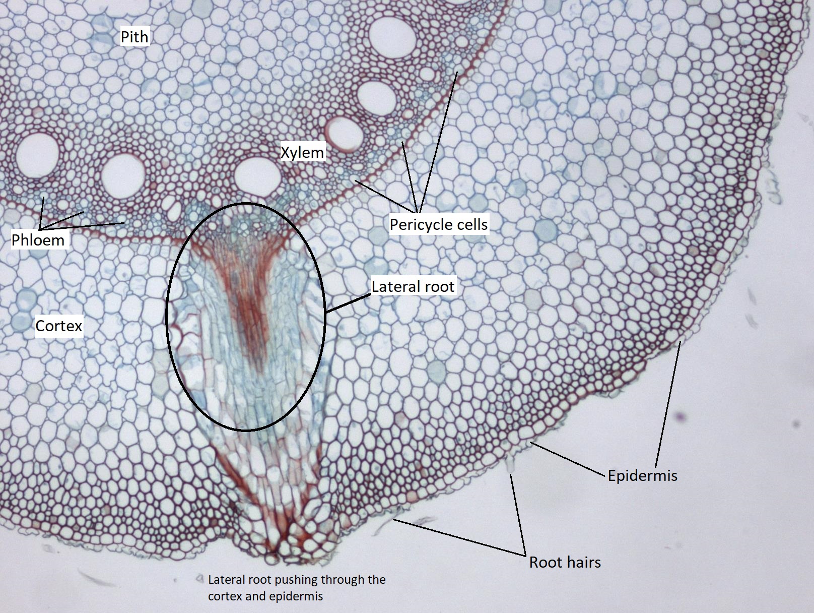
Most of the root is composed of cortex tissue, and the endodermis, the innermost layer of the cortex, borders the stele. The outer layer of the root (external to the cortex) is the epidermis. Figure 3.2.3. 4: Eudicot root cross section. From center out, the xylem (in red) make an X, and the side tissues (green) make up the phloem..

monocot stem anatomy
Aim: To prepare temporary stained glycerine mounts of transverse sections of stem and root of Dicot and Monocot plants. Material required: Sharp razor, brush, dropper, needles, watch glass, microscopic slides, cover-slips, safrannin, glycerine and compound microscope. Technique: Take 2-3cm long pieces of the material.

Transverse section of monocot root Biology diagrams, Root, How to plan
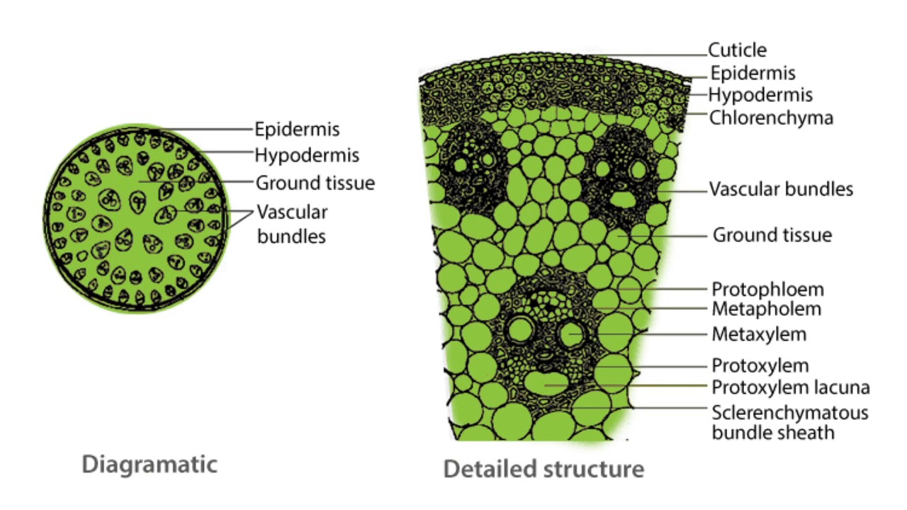
The transverse section of the sunflower root is depicted in the diagram. The following is the internal tissue organization:. In the transverse section of a monocot stem, the epidermis, hypodermis, and ground material are distinguished. The monocot stem's interior tissues are arranged in no particular order.
NCERT Solutions for Class 11th Ch 6 Anatomy of Flowering Plants Biology « Study Rankers
Transverse Section of the Monocot Root Transverse Section of the Monocot Stem Things to Remember Previous Year Questions Sample Questions Key Terms: Monocot Root, Monocot Stem, Dicot Root, Dicot Stem, Transverse Section Epidermis, Vascular Bundles, Pith, Cortex Aim [Click Here for Sample Questions]
Preparation and Study of Transverse Section of Monocot and Dicot Roots and Stems
Zea mays. Zea mays (corn) is often used as a model organism for monocot anatomy. Figure 11.1.2.1 11.1.2. 1: The images above show a corn seedling in two different stages of development. The first image is of the corn seedling at an earlier stage. It has produced a shoot (with one cotyledon) and a long root (radicle).

Transverse section of a part of a typical monocot root has been sho... YouTube
Aim To prepare a temporary stained mount of a transverse section of dicot and monocot stem and root to study various plant tissues. Material Required Preserved material of sunflower root and stem Preserved or fresh material of maize root and stem Microscope Sharp blade Slides Watch glass Coverslips Safranin (1gm in 100ml of 50% ethanol) Brush

Monocot Stem Cross Section
Transverse section of a part of a typical monocot root has been shown in the given figure. Identify the different parts (from \ ( A \) to \ ( E \) ) and select the correct option. (a).
Monocot Stem Cross Section Monocot Root Cross Section Structure (with PPT The main
Overview Test Series Objective The goal is to create a temporary stained mount of a transverse section of dicot and monocot stem and root to investigate various plant tissues. Crack SSC CHSL with India's Super Teachers Get 6 Months SuperCoaching @ just ₹489 ₹449 Your Total Savings ₹40 Purchase Now Want to know more about this Super Coaching ?
11.1.2 Monocot Roots Biology LibreTexts
Monocot root In monocot roots, the vascular bundles are arranged in a circular pattern. Monocots and dicots contain two main types of vascular tissue: the xylem and phloem. The xylem carries water and dissolved minerals upward from the roots to the stem and leaves.